Seeing Inside The Eye
Improvements in laser scanning technology reveal the intricacies hidden inside our eyeballs.

Image by Peter Maloca, courtesy of Wellcome Library, London
Place yourself in the image above, and you might think you were in the depths of the earth.
In reality, you’re looking at the depths of a healthy human eye—specifically, a spot that’s about the size of 100 microns (or about 1/10th the thickness of a fingernail). Ophthalmologist and eye surgeon Peter Maloca created the image using an advanced imaging technique called swept-source optical coherence tomography, which can be used to probe the eye and retrieve detailed scans in mere seconds.
Optical coherence tomography (OCT) “works like ultrasound, but instead of sound waves, we use laser light,” says Maloca, who is also a post-doc at the University of Basel’s Department of Ophthalmology in Switzerland. The laser used in swept-source OCT is only administered for short amounts of time, so it’s not destructive to tissue. A camera-like device directs the laser to the eye in pulsed waves, and the light that’s reflected back offers enough data for researchers to subsequently create penetrative, 3-D images—like the one above—of structures inside the eye. To sharpen borders, Maloca and his team developed a method of post-processing that enables them to remove artifacts that cause visual noise.
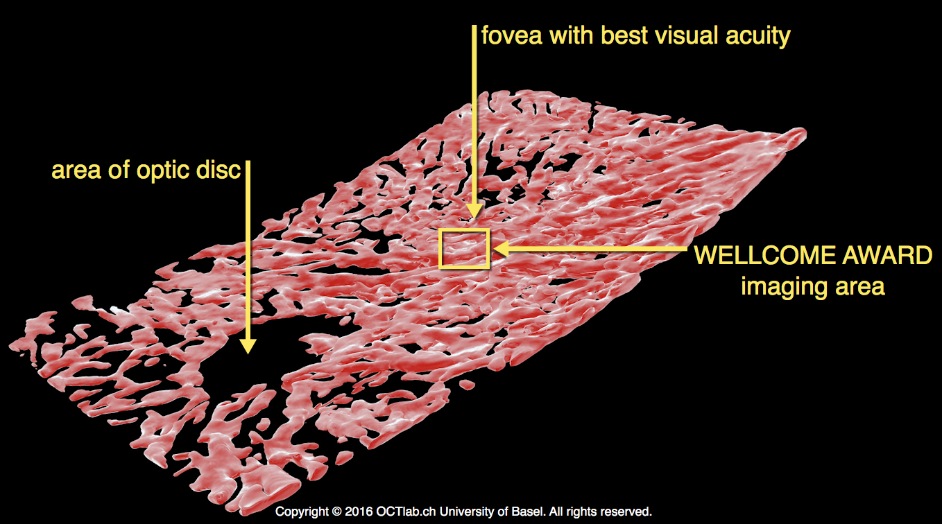
This particular image, which was a winner in this year’s Wellcome Image Awards, shows the inside of tiny blood vessels that provide oxygen and nutrients to a region of the retina called the fovea. Humans, Maloca explains, have two kinds of vision: central vision that is used for seeing in detail, such as when you read, and peripheral vision, which is used for orientation, like when you’re walking. The fovea is responsible for the sharpest central vision.
“If you have changes there, you are losing your visual acuity,” Maloca says.
The award-winning image “looks like a cave, because normally you have blood cells inside that space, but the blood cells move so fast that you actually can’t image them,” Maloca says. “The space is empty because we can’t track them with the laser scanner.”
Maloca chose the colors and textures and added them in later on a computer to highlight areas of interest. “In the upper part, where the more-red light is—that is the retina, so you are really in the bottom of the eye,” he says. The blue color represents the outer layers of the eye. “In the center, the illuminated center, there is a statue-like structure,” he says, which his team thinks might be involved in the regulation of blood flow. They’re researching this mysterious structure to confirm its role.
Maloca hopes to use this technology to better identify and track diseases in the eye. “For example, in patients who have diabetes, when you look inside the eye [using standard techniques], it’s sometimes difficult to establish the stage of this disease,” he says (retinal tumors can develop in people with diabetes). “Then there are patients who have age-related macular degeneration. This is one of the most common diseases; this is destroying central vision. [With this technology,] we hope to make an early detection of changes in the retina.”
In the event that you want to journey into the cavernous eye, Maloca has helped develop a game to display how advanced OCT technology has become. Go for a ride here.
This article was updated on May 9, 2016, to include an updated version of the 3D model image.
Chau Tu is an associate editor at Slate Plus. She was formerly Science Friday’s story producer/reporter.