The Hidden Beauty Of The Human Placenta
Though discarded after birth, the placenta builds the first vital connection between mother and fetus.
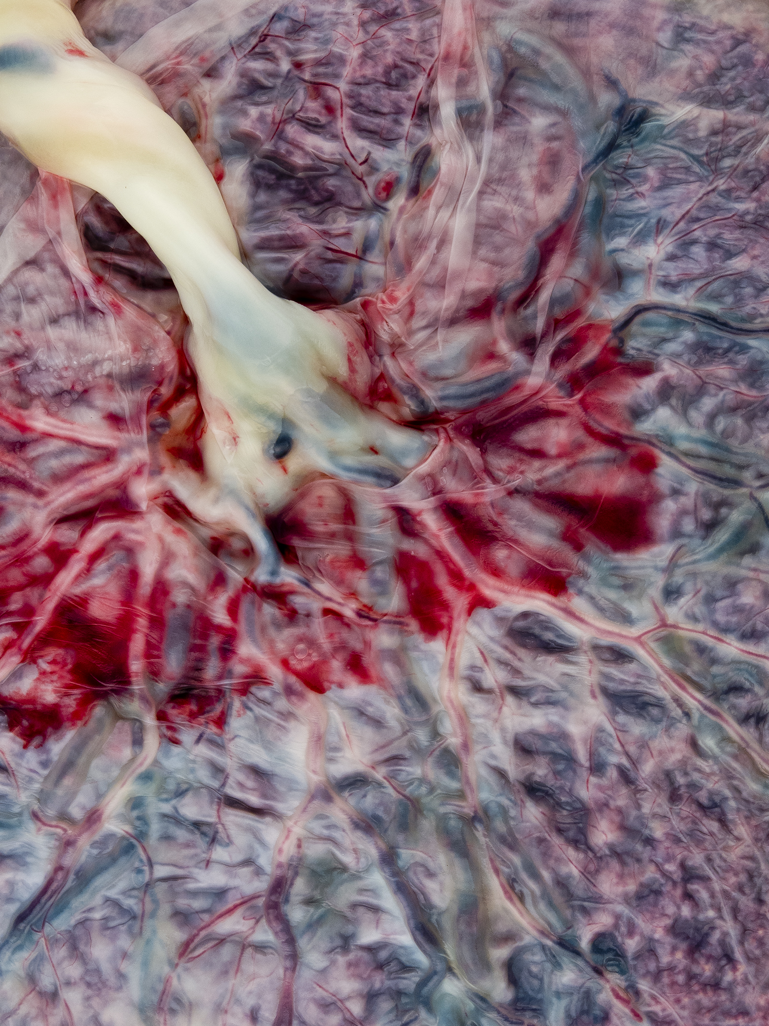
Arteries and veins fan out from the umbilical cord across the placenta, seen through the thin film-like covering of the amniotic sac, left behind after birth. The dark spot at the center of the image isn't an abnormality; it shows a particularly thick, dark blue vein emerging from the cord. Credit: © Norman Barker, Johns Hopkins School of Medicine
What you’re seeing is a close-up of the human placenta, pictured from the side that would face a fetus in the womb. The twisting white branch is the umbilical cord.
“If somebody didn’t know what a placenta looked like, they might think this is a landscape with its rivers and tributaries,” says Christine Iacobuzio-Donahue, a pathologist from the Memorial Sloan Kettering Cancer Center in New York City. She collaborated with Johns Hopkins School of Medicine medical photographer Norman Barker to highlight these striking abstract patterns for Hidden Beauty, their book of medical art photography. This particular placenta had just arrived from the delivery room at Johns Hopkins Hospital.
Iacobuzio-Donahue and Barker thought the fetal underside of the organ showed a more compelling face than the upper half. “The maternal side looks very ragged, very beefy. There’s definitely nothing attractive about it at all,” says Iacobuzio-Donahue. But on the fetal side, the remains of the amniotic sac, left clinging to the placenta after birth like a burst balloon, form a fine gauze over the tissue, she explains. “That’s why it’s so smooth and pearlescent-looking.”
Barker zoomed in on this 15 cm x 25 cm section of the placenta, revealing blue-black veins and scarlet arteries extending from the white umbilical cord. In the womb, this dense network of vessels roots in the placenta’s vascular tissue, where a vital transaction takes place between the mother’s blood and the fetus’s through diffusion (the blood never mixes directly).
Nutrients and oxygen from the mother’s blood vessels feed into the placenta, where the foot-long umbilical cord channels the precious cargo through an internal vein to the awaiting fetus. In exchange, two arteries carry fetal waste like carbon dioxide back through the cord to the placenta to be transferred to the mother for disposal via her bloodstream. The organ also plays a role in guarding the fetus against bacterial infection, and transfers hormones that control the mother’s metabolism and aid the baby’s growth.
This intricate support system grows anew along the lining of the uterus whenever a fetus starts to develop, only to be expelled completely after the baby is born. But before they remove the organ, pathologists use it to crosscheck the health of mother and child.
“Things they look for are that the umbilical cord has a normal number of vessels—two arteries and one vein; that the vessels of the placental disk are normally developed; and that there aren’t any cellular changes to suggest a viral infection,” Iacobuzio-Donahue says. Thicker placentas can indicate diabetes in the mother, and slimmer organs could mean a baby is weak and needs extra care. (This placenta above was healthy.)
The picture draws attention to an organ that, while ephemeral, once sustained us all. “The placenta really is a marvel of design,” says Barker. “It is the only organ designed to be disposed of after it performs its function.”
This post was updated on March 5, 2015, to reflect the following corrects. A diagram that we linked to contained several errors, so we removed it. Also, the article originally stated that the umbilical cord channels nutrients and oxygen “through two internal arteries to the awaiting fetus. In exchange, a single vein carries fetal waste like carbon dioxide back through the cord to the placenta to be transferred to the mother for disposal via her bloodstream.” In fact, a single vein carries nutrients and oxygen to the fetus, and two arteries carry fetal waste back to the placenta.
Emma Bryce is a freelance science writer whose articles have appeared in publications such as The Guardian and Audubon.