Practice Eye of Yesteryear
The instrument is an early version of a training device popularized by optometrists.
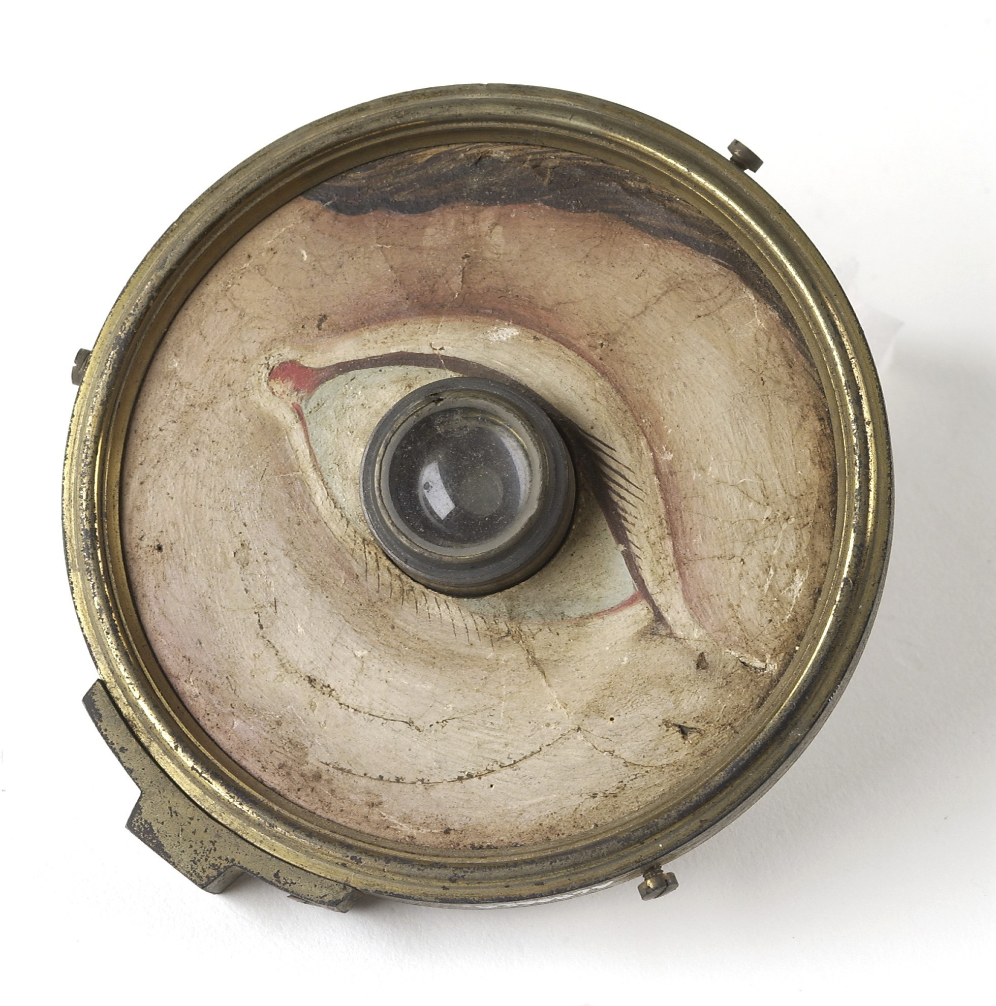
Practice eye, with brass-backed paper front with hand-painted face around eye, by W. & S. Jones, London. Credit: Wellcome Library, London/CC BY-NC-ND 4.0
In 1800s London, if you needed an optical device—eyeglasses, periscopic spectacles, a microscope—W. & S. Jones was the place to go. Founded by two enterprising brothers, the company specialized in various scientific instruments, like the model eye above. It could be used to demonstrate—at a very basic level—how the eye refracts light.
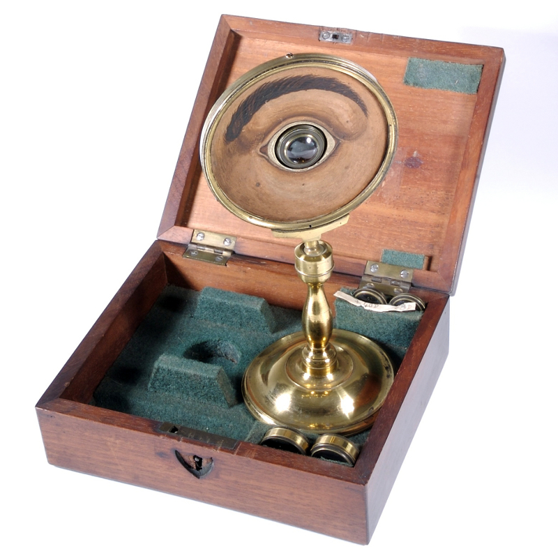
The instrument is an early version of a “practice eye,” a training device that became more popular in the early 20th century, when optometry emerged as a field associated with healthcare. In the Romantic era, however, “optician” translated to “a skilled craftsperson who makes things using scientific and physical principles,” says Neil Handley, curator at the British Optical Association Museum, part of London’s College of Optometrists. W. & S. Jones’s practice eye was “an idea that was way ahead of its time and was subsequently put to very practical use.”

The part that looks like a fish’s bulbous orb consists of several parts that simulate structures in the eye. An outer piece of glass represents the cornea, followed by an artificial “iris” supported by another glass insert. Next comes a small glass disk—the “lens”—and then a thicker, translucent chunk to symbolize the eye’s vitreous jelly. The back of that chunk also stands in for the retina, sealing everything off on the other side. Meanwhile, hand-painted facial features, including an eyebrow and eyelashes, frame the assemblage, lending a touch of beauty, according to Handley.
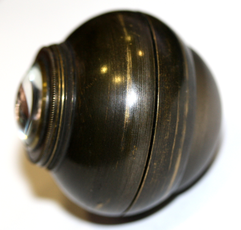
The instrument came with interchangeable glass corneas that could be switched out to simulate different corneal curvatures. A few “corneal caps” also accompanied the device, which could be screwed individually on top of the cornea to mimic a spectacle lens. Because the corneas and corneal caps had different powers, they refracted light to varying degrees.
Written instructions that might have existed have been lost to time, but Handley suggests that to use the model eye, someone would have shone a pencil beam of light—perhaps created with a candle held up to a pinhole pricked through a piece of cardstock—on the cornea and observed how the beam refracted onto the retina depending on the power of the cornea or corneal cap. “It’s a very simple means of demonstrating that eyes of different focal lengths will see in different ways,” says Handley.
Although the instrument could have been used to teach doctors the simple principle of light refraction in the eye, it likely served another purpose, too. Just as someone might collect classic novels to give the impression of being well read, Romantics with a scientific bent may have kept a model eye at home to “impress their friends and make them think that they’re part of this scientific revolution, that they are learned people,” says Handley. “These are beautiful objects for aesthetic, decorative purposes in addition to any educational benefit they might convey.”
Julie Leibach is a freelance science journalist and the former managing editor of online content for Science Friday.