Gift Of The Mummy
A patient more than 3,000 years-old takes a turn through a CT scanner.

The first two mummies arrived at Barnes-Jewish Hospital in St. Louis, Missouri, at 7:30 a.m. on a cold, gray, and rainy Sunday this October. A third arrived a bit later. By 12:30 p.m., all had taken a turn through a CT scanner.
Four radiologists, each interested in different parts of the body, are scrutinizing the scans for clues into the mummies’ lives, medical conditions, and deaths. Their discoveries will add context to the St. Louis Art Museum’s Egypt collection, planned for a reinstallation in 2016. (Hear more about the mummies on this week’s show, airing on Halloween.)
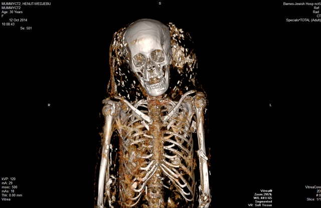
The individual pictured above, known as Henut-Wedjebu, is the oldest and least examined of the three. “The challenge was scanning a patient that just happened to be from 1300 BC,” says Sanjeev Bhalla, the project’s lead radiologist, who usually tends to living patients.
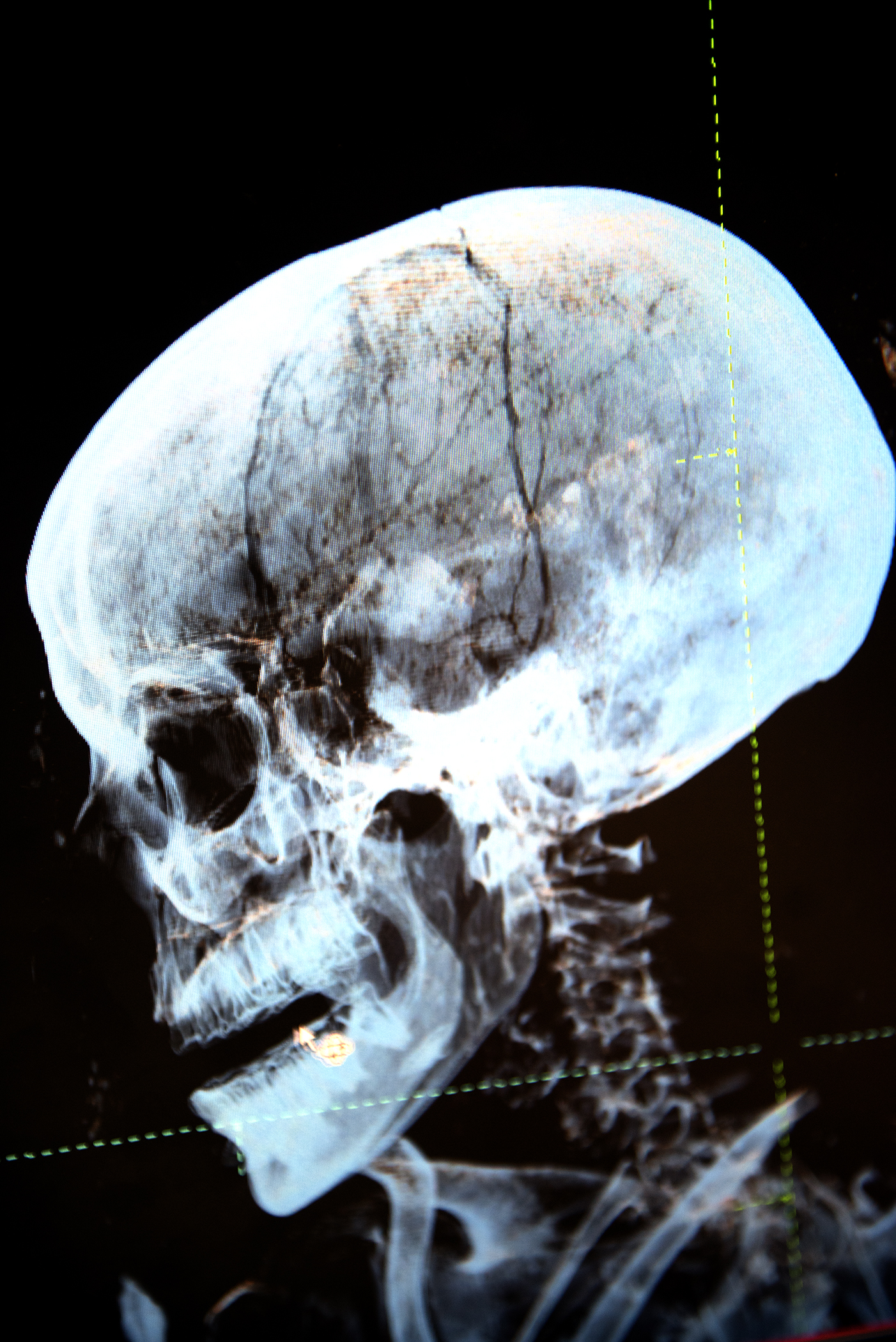
An x-ray taken almost 50 years ago showed a fracture in Henut-Wedjebu’s skull, but the radiologists knew little else about her physical state. Computed tomography probes deeper than conventional x-ray imaging, providing more detail and contrast and picking up on calcified structures filled with gas.

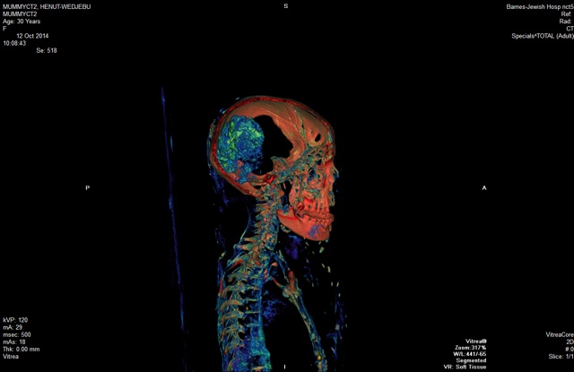
“You can see her optic nerve, you can see her spinal cord,” says Lisa Çakmak, assistant curator of ancient art at the St. Louis Art Museum. “The level of anatomical detail you can see is stunning.”
The ornateness and size of Henut-Wedjebu’s gold-foiled coffin, as well as an inscription that translates to “Singer of Amun and Lady of the House,” suggest that she was a noblewoman. The CT scans offer more personal insight—for instance, she was mummified with a halo of glass-looking beads, perhaps signifying that her hair was decorated or that she wore a headdress.
Henut-Wedjebu’s facial bones also show up “phenomenally well” in the scans, says Bhalla. He suspects that she was beautiful, with a slender face and high cheekbones. And her remaining teeth are still in good condition.
After locating and studying the skull fracture, the team suggests that if the injury happened before death, it would have been traumatic. The doctors spotted signs of infection as well, such as calcification in the lymph nodes and scarring in the lungs’ lining—another possible cause of death.
Scanning also showed a shrunken but intact brain. Indeed, Henut-Wedjebu was mummified during a rare pocket of ancient Egyptian history when brains were left in. (Her male counterparts, 1,000-1,100 years her junior, are missing their brains.)
Other features, distorted by dehydration, were less discernable. “We’re still poring through those images to make sure we can properly identify organs,” says Bhalla. For instance, they’re continuing to search for Henut-Wedjebu’s heart, as well as those of her fellow mummies.

The radiologists are decoding the mummies’ scans separately and will reconvene around December to finalize their observations. That way, they’ll each have the chance to form independent opinions about what the images signify.
The experience has provided lessons beyond history and anatomy. “The success of the project really required us to go back to the fundamentals of learning how to be a doctor,” says Bhalla, such as treating patients and their families (in this case, art institutions) respectfully, building on technological experience, and puzzling out available data.
“In the end,” says Bhalla, “this so-called curse of the mummy may have been the gift of the mummy.”