Mapping Brain Connections Reinforces Theories On Human Cognition
17:12 minutes
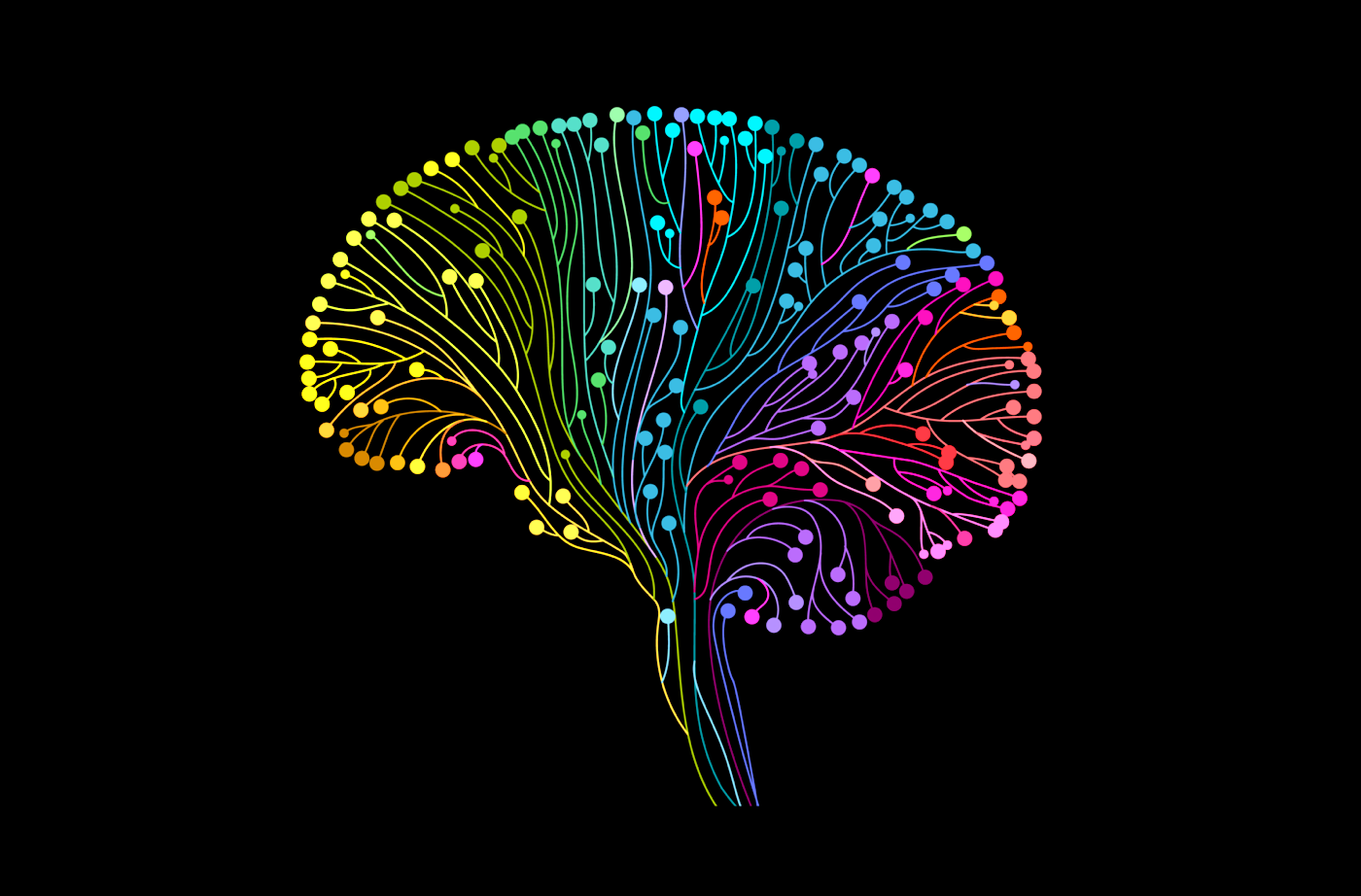
Brain regions are associated with different functions—the hippocampus is responsible for long-term memory, for example, and the frontal lobe for personality, behavior, and emotions.
After decades of research using sophisticated brain imaging, there’s a growing consensus among neuroscientists that understanding the connections between brain regions may be even more important than the functions of the regions themselves. When it comes to understanding human cognition, the whole is greater than the sum of its parts.
Ira speaks with Dr. Stephanie Forkel, assistant professor at the Donders Centre for Cognitive Neuroimaging at Radboud University in Nijmegen in the Netherlands, who wrote a review article in the journal Science about the importance of brain connectivity, and what it means for the future of neuroscience.
Invest in quality science journalism by making a donation to Science Friday.
IRA FLATOW: This is Science Friday. I’m Ira Flatow.
For the rest of the hour, we’ll be focusing on a paradigm shift in the field of neuroscience. Your standard textbook maps out the brain into regions of the body. You’re hearing is here, your left leg there, the hippocampus responsible for long-term memory, the frontal lobe responsible for your personality, behavior, and emotions, right? But now, after decades of research, using precision brain imaging, there’s a growing consensus among neuroscientists that understanding the connections between brain regions may be even more important than the regions themselves. Basically, the whole is greater than the sum of its parts.
My next guest recently wrote a review article in the journal Science about the importance of brain connectivity and what it means for the future of neuroscience. Dr. Stephanie Forkel is an assistant professor at the Donders Centre for Cognitive Neuroimaging at Radboud University, based in Nijmegen, in The Netherlands. Welcome to Science Friday.
STEPHANIE FORKEL: Hello.
IRA FLATOW: Nice to have you. How much of a paradigm shift is going on here? I mean, haven’t neuroscientists known that brain connections were really important for decades? Patients who have had brain injuries in one part of the brain, other parts of the brain compensate for that. Why now are we closing in on this brain connectivity model?
STEPHANIE FORKEL: So you’re absolutely right. We knew for decades, or even centuries, that we had those white matter connections, which are basically the superhighways of signal propagation in the brain and they connect different parts of the brain. But only now do we actually have the large enough data sets and new tools to measure what those white matter connections actually do and what they connect in the living human brain.
IRA FLATOW: So should we now throw out those old models of brain regions, or is that still part of the equation?
STEPHANIE FORKEL: That’s still part of the equation. So we’re not going from one extreme to the other. And the truth is somewhere in the middle. So you still need those brain areas. I mean, there’s a reason we have the cortex of the brain– so the surface areas. But an area in itself can’t really perform all the functions that we know we can do. So if we take the example of language, there is a part in the frontal lobe that’s important for articulation, and there’s a part of the brain in the temporal lobes– so close to your ears– that is important for understanding language.
Now, if we talk in a normal conversation, we need both of these areas to react very quickly and dynamically in a conversation. And this dynamic interaction of areas is facilitated by the white matter connections.
IRA FLATOW: So those are the connections you’re talking about, something called white matter. What is that? What’s the difference between white matter and, let’s say, something else we hear about in the brain, gray matter?
STEPHANIE FORKEL: So generally speaking, the gray matter is the surface of the brain, the outside. So when we talk about the brain, the picture that comes to your mind of that classical half a hemisphere, looking at the brain from the outside, that’s the gray matter. And the gray matter is composed of brain cells. So those neurons are in the gray matter.
And then we have the white matter, which is the connective tissue. So all those brain connections, the highways as I said, that connect those different parts of the cortex together.
IRA FLATOW: They must be pretty fast connections. I mean, they must be able to talk quickly with each other, right?
STEPHANIE FORKEL: Yes, absolutely. So it’s estimated that the speed of signal conduction along the white matter is about 300 kilometers an hour, which is equivalent to a bullet high-speed train.
IRA FLATOW: Wow. Did we always know that, or did it take modern brain imaging to figure that out?
STEPHANIE FORKEL: Well, that you actually would figure out with electrophysiology. So you actually can follow the electrical signals that are sent along the white matter to measure the speed. So you can basically input in one part of the brain and see how long it takes for it to be received at the other end of the brain.
IRA FLATOW: But you use other types of imaging, don’t you, to do your work and to study the brain?
STEPHANIE FORKEL: Correct. So we use a method called diffusion-weighted imaging tractography, which is based on an MRI scanner– so that big tube that many people probably are familiar with– to look inside the living human brain and look at the structure of those connections. So not so much the function that you would get when you measure the speed of conduction, but really the anatomy of those connections, where they are, what they connect to.
IRA FLATOW: Are you putting living, breathing people in this MRI, or what else might you be doing?
STEPHANIE FORKEL: Yes, we do most of the time. Not all the time. But ideally, we would imagine living, breathing people in the MRI scanner. And it’s completely noninvasive. So you can go in there. You can even have a nap whilst we take those pictures. So for some people, it’s quite a pleasant environment.
IRA FLATOW: Wow. I cannot imagine that. I’ve had a brain scan, an MRI. It is not a pleasant situation. Well, I have claustrophobia. So that would do it, right?
STEPHANIE FORKEL: Yeah, that doesn’t help.
IRA FLATOW: But you also look at postmortem brains, too, right?
STEPHANIE FORKEL: Correct. So we look at postmortem brains sometimes in the MRI scanner. But we also actually look at them in the morgue and do actual Klingler postmortem dissections. And that lets you really carve out the white matter connections between those brain areas in the actual brain.
So when we use the diffusion-weighted imaging tractography, it’s a proxy of the anatomy because it’s based on how water molecules move around in the brain. And if you have areas where there is no restriction– so for example, there’s these holes, basically, in the brain that are filled with liquid to cushion the brain when we walk or hit our head– then the water molecules can freely move around in all different directions and there’s no restriction.
The water molecules within those white matter connections, however, they’re limited by the surface of those connections. So it’s basically like a straw. And they travel faster along the direction of the axons– so that connection– and they’re hindered in the perpendicular direction. And this is what we actually pick up with the MRI machine. So it’s an indirect estimate of where those connections are. Whereas, Klingler postmortem dissection actually lets you see them in real life.
IRA FLATOW: How did you discover– or how did people discover– that this technique you’re using would be a good way of studying the connections in the brain?
STEPHANIE FORKEL: So there’s this famous saying that someone’s noise is someone else’s signal. And what that means is that when you acquire images in an MRI machine, there is a lot of noise in the data. And for many years, we thought we need to get rid of this noise. It’s disturbing the signal that we’re interested in, which is the anatomy.
And then people started realizing that actually within this noise that is in the images there is some patterns. There some information that we could actually use and study. And this is how diffusion tractography was invented, or resting state imaging these days, for example.
IRA FLATOW: Yeah. That reminds me, in physics, in astronomy, of accidentally discovering the cosmic background radiation because they thought there was noise in the receiver. So this is an interesting parallel.
STEPHANIE FORKEL: Yeah, exactly.
IRA FLATOW: The idea of defining the functions of brain regions, let’s talk about how old that is, right. Where did it come from?
STEPHANIE FORKEL: So defining the function of brain regions is actually a very old way of doing clinical anatomical correlation studies. And that really comes from seeing patients in the beginning. So we have a series of famous cases to actually establish the neurosciences as we know them today. And that is because those famous cases had a damage to the brain.
So quite famous, for example, is Phineas Gage, who had an iron rod that flew through his skull and his brain, and then landed a couple of meters behind him. And that iron rod damaged a part of his brain in the frontal lobe. He could still talk, but what had changed was his personality, his inhibition. So that is one of the famous cases that placed personality and so-called executive functions in the frontal lobe.
Another famous case is Patient Tan. And he’s known as Patient Tan because he suffered a stroke to the frontal lobe, again, a different part, and he could only utter the word “tan.” He was French, and hence the slight imitation of an accent here. And that placed the articulation of language in the posterior frontal lobe.
And what these patients allowed people to do is to look at the brain and look at what function the patient lost. And that was then put together as in, if you lose the function of the damage to this part of the brain, then this part of the brain is responsible for this kind of function. And this is where this area to function mapping originated.
IRA FLATOW: Quite interesting. Can you also, using this model, help us better understand how the human brain evolved?
STEPHANIE FORKEL: Yes, absolutely. So as we said before, ideally, we put living, breathing humans in the MRI scanner, but we don’t do this all the time. So you can also put non-human primates in the scanner. For example, macaques or sometimes even chimps. And that lets us study the white matter in different species. And then, by proxy, you can make deductions about when the white matter changed between the species. And that would give you an indication of when certain cognitive functions may have evolved in the human brain.
IRA FLATOW: Such as language?
STEPHANIE FORKEL: Such as language, for example, yes.
IRA FLATOW: So some monkeys may have some but not all the connections in the brain, so to speak, that we have?
STEPHANIE FORKEL: Correct. So there’s a very prominent language connection that we call the arcuate fasciculus, which means the arching fiber bundle. And you can see a precursor of that in the monkey brain, but it’s not the full connection. So it doesn’t reach exactly the same cortical areas. And it’s not as prominent as it is in the human brain, for example.
IRA FLATOW: Of course, everyone’s brain is a little bit different, right? How can this networking idea help you understand the individual differences between the brains– all of our brains?
STEPHANIE FORKEL: So what we have done for decades of neuroimaging is that we wanted to know how the brain works. What’s the function and the structure of the typical brain? And in order to do that, you take a picture of a lot of individual brains, and then you bring them all together in a reference space where they all look the same. So you then compare them across the group.
Now, what we can do nowadays, because we have a lot more data available and we have new analysis tools available, we can actually look a lot more into detail into each and every single individual brain. And what we have done, back in 2008 for example, is look at the variability of the white matter. And what we could see is that there is a gradient in the brain, whereby older parts of the brain tend to be less variable, meaning they’re more alike between us, and newer parts of the brain and the areas that are related to cognitive functions, using functional imaging studies or lesion studies, were actually highlighted as being more variable. So there’s this beautiful gradient of variability in the brain.
IRA FLATOW: Yes. So would it be possible then to map how, say, a mental health issue or someone’s personality changes the structure of the brain?
STEPHANIE FORKEL: To a certain extent, yes. So there’s already group studies that would compare people with certain neurological or psychiatric disorders to so-called healthy controls and see what is different in those brains. What you can also do is look at clinical populations– again, this could be neurological or psychiatric patients– and follow them over time, so in a longitudinal study design, and see how the brain– that could be the gray matter or the white matter– changes throughout time.
IRA FLATOW: This is Science Friday, from WNYC Studios. In case you’re just joining us, I’m talking with my guest Dr. Stephanie Forkel, about the importance of understanding how brain regions are connected.
Now, of course, we have those two hemispheres of the brain, as you mentioned. What can looking at these connections help us better understand about the functioning– and possibly the different functionings– of the left and right brain?
STEPHANIE FORKEL: So there’s this conception that we have a left brain and a right brain and the functions are dramatically different. So classically speaking, the left hemisphere is important for language and the right hemisphere is more important for things like visual, spatial attention, for example.
Now, one thing that we need to consider is that those two hemispheres are densely connected by white matter. So they’re not freely floating around in our skull, but they’re actually densely connected. And every part of the left hemisphere is connected to every part in the right hemisphere pretty much. And that means that some of the functions can be traced in both hemispheres.
Now, obviously, the way we look at function these days– for example, language, we often look at the production and the comprehension of language. But language is a lot more, right? There’s intonation. There’s prosody. There’s understanding humor, for example. Or when we think about idioms, you understand the meta meaning of the words. And some of those functions are actually performed by the right hemisphere and not the left hemisphere.
So what we sometimes see, for example, is that patients who suffer a lesion to the right hemisphere, they can articulate themselves, they can understand language, but when they talk it’s very monotonous. So this clear-cut division of this is what the left does and this is what the right does is not really appropriate. And also, when it comes to recovering from brain damage, we can see that there’s also some dynamic interaction between the left and the right trying to recover some of the function.
IRA FLATOW: So how can your network model help scientists come up with better treatments for people with brain injuries or neurodegenerative disorders?
STEPHANIE FORKEL: So what we have seen in clinical studies is that when you only look at the gray matter lesions– so where the lesion is located on the surface of the brain– we can explain some of the variance that we see in the recovery of people, but not all of it. Whereas, if we look at the networks that are actually disconnected by a lesion, that explanationary power increases dramatically. And we still cannot explain all of the recovery that we see, but at least a larger part than just looking at the lesion location itself.
IRA FLATOW: Let’s talk about what the next big question– I’m always interested in where you’re heading with your research– what’s the next big question you’re looking to understand about the brain?
STEPHANIE FORKEL: So in my own research, I’m currently trying to understand the magnitude of variability. So it’s well established that our brains are different. And we have beautiful maps showing the variability of certain parts of the brain. And we, as I mentioned, did this study in 2018, where we looked at the variability at the group level. But I’m really trying to bring this to the individual brain now and map the magnitude of variability and what that magnitude then means for our cognition and health and in disease.
IRA FLATOW: Do you think it’s possible the brain– it’s got, what, how many billions and billions of cells in it– is it possible to understand our brain, do you think, completely, and even get to consciousness some day?
STEPHANIE FORKEL: The Holy Grail.
IRA FLATOW: I had to ask that question.
STEPHANIE FORKEL: I think I’d be surprised if we managed to get there during my career, but it it’s worth trying.
IRA FLATOW: Good answer. Thank you for taking time to be with us today.
STEPHANIE FORKEL: My pleasure. Thank you.
IRA FLATOW: Dr. Stephanie Forkel, assistant professor at the Donders Centre for Cognitive Neuroimaging at Radboud University, based in Nijmegen, in The Netherlands.
Copyright © 2022 Science Friday Initiative. All rights reserved. Science Friday transcripts are produced on a tight deadline by 3Play Media. Fidelity to the original aired/published audio or video file might vary, and text might be updated or amended in the future. For the authoritative record of Science Friday’s programming, please visit the original aired/published recording. For terms of use and more information, visit our policies pages at http://www.sciencefriday.com/about/policies/.
Shoshannah Buxbaum is a producer for Science Friday. She’s particularly drawn to stories about health, psychology, and the environment. She’s a proud New Jersey native and will happily share her opinions on why the state is deserving of a little more love.
Ira Flatow is the founder and host of Science Friday. His green thumb has revived many an office plant at death’s door.