Subscribe to Science Friday
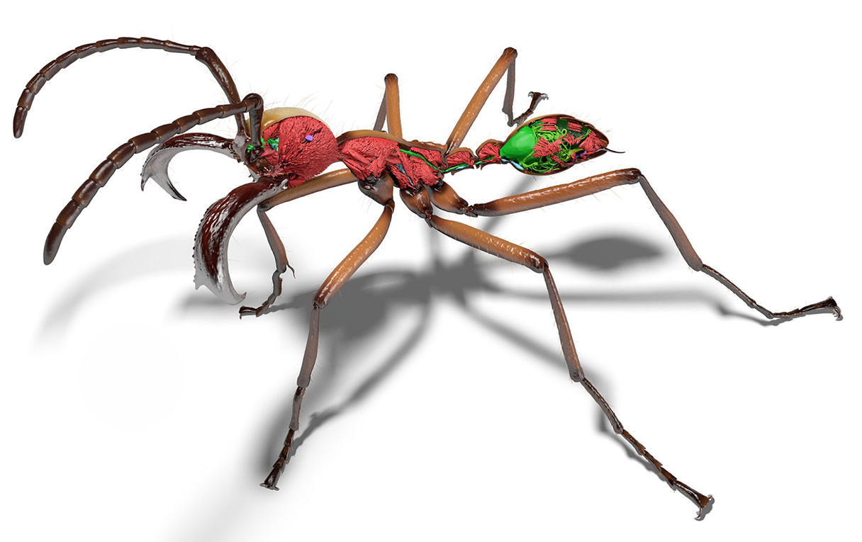
A project called Antscan has generated high resolution images of thousands of ants, representing over 700 species. To make it happen, researchers brought preserved ants from collections around the world to a particle accelerator in Germany. There, a powerful synchrotron x-ray source combined with a vial-swapping robot allowed the researchers to build a collection of 3D ant images, inside and out. Each voxel (like a 3D pixel) has a resolution of 1.22 micrometers—enough to see the tiny hairs on ant bodies, and distinguish individual muscle fibers.
Antscan researcher Julian Katzke joins us to describe the background of the project, and how the images could be used for science and art.
Check out images from the Antscan projet:
Donate To Science Friday
Invest in quality science journalism by making a donation to Science Friday.
Segment Guests
Julian Katzke
Dr. Julian Katzke is a postdoc at the Smithsonian National Museum of Natural History. He worked on the AntScan project while a PhD student at the Okinawa Institute of Science and Technology.
Segment Transcript
[MUSIC PLAYING] FLORA LICHTMAN: Hi, I’m Flora Lichtman. And you’re listening to Science Friday. In the seminal insect film A Bug’s Life, we’re given a bug’s eye view of the world.
SPEAKER 1: Now, stay calm. We are going around the leaf.
SPEAKER 2: Around the leaf? I don’t think we can do that.
SPEAKER 1: Oh, nonsense. This is nothing compared to the twig of ’93.
FLORA LICHTMAN: We come face to mandible with ants, their antenna, their spindly legs, everything. And they’re delightful. But perhaps the accuracy left a little something to be desired. Enter the Ant Scan Project, which has generated high-resolution X-ray images of over 2,000 real ants from over 700 species. And I gotta say, the results really are stunning. This is ants in all of their beautiful and frightening glory.
Joining me now to dig into this mound of ant data is Dr. Julian Katzke. He worked on this project while a PhD student at the Okinawa Institute of Science and Technology. Hi, Julian.
JULIAN KATZKE: Hi, Flora. Thanks for having me.
FLORA LICHTMAN: Thanks for being here. OK, so you came into this work comparing the mouthparts of dozens of ant species. Why mouthparts?
JULIAN KATZKE: Oh, so if you’re a human and you live anywhere in the world, there’s a pretty high chance that ants are all around you. And there’s this now pretty well-known fact that the biomass of all ants equals or surpasses that of all humans. And the total diversity of ant species is enormous. And that also extends into their forms and shapes, particularly in the mouthparts, because they are the first tool that ants use in their daily lives.
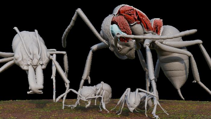
So our mascot model is this South American army ant. They have small ant workers, but then also large soldiers. And these large soldiers have these fishhook-like mandibles that you could even use to staple a wound. Their bite is so fierce that they would never let go. And I think the theory here is that these mandibles are really just there to hurt something that is as big as a human. And so really learning more about the evolution of these different mouthparts was motivating my PhD research.
FLORA LICHTMAN: Describe for people who can’t see it. And we’ll put some on our website at sciencefriday.com/ants. What do the images look like?
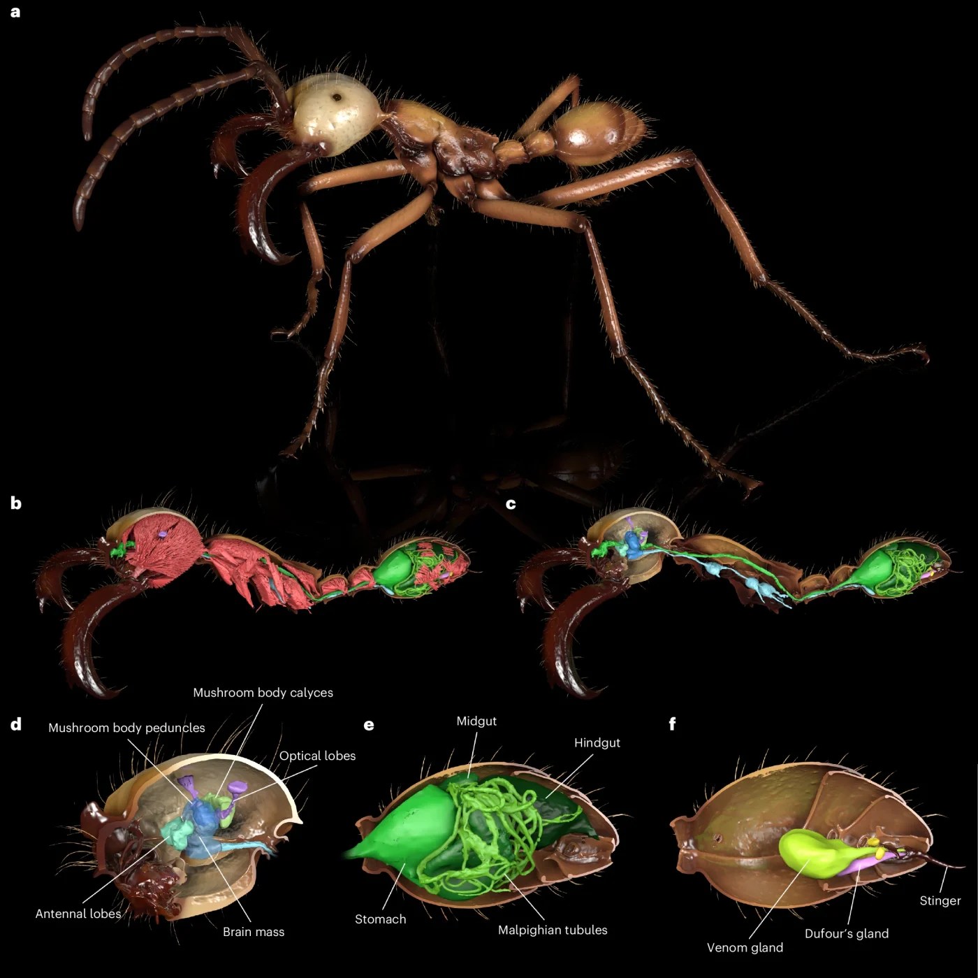
JULIAN KATZKE: So the images, they are derived from X-ray images. So if you’ve ever taken an X-ray image at a hospital, you have this see-through X-ray image. And then if you would turn yourself around while taking these X-ray images enough times, you could mathematically reconstruct the 3D volume. So that is the technology behind computer tomography. And that’s exactly what we did with ant scan. The data that we end up with are like slices of images that are like grayscale. And so they contain the anatomy of the ants.
FLORA LICHTMAN: And then you put them together, and you can color them and make them beautiful, too.
JULIAN KATZKE: Yes. So yeah. So the tomography data is like the raw data. And there’s plenty of methods that you can use to generate something out of it. So in the first instance, it might just be like a measurement, like body size or whatever. But you can also use them in a way like Hollywood 3D animators would.
FLORA LICHTMAN: Yeah, I mean, that’s what caught my eye. They were just amazing. They’re like 3D, but in this extreme detail. And they look like aliens.
JULIAN KATZKE: I mean, the ants only look like aliens because they look alien to us. They are a bit strange because they’re so small, and our eyes are just not good enough to really take it all in at once. And I think it’s one of the basic aspects why a digital library of animal shapes and forms can be very important, is because so much of the world out there is just so tiny. And for us to engage with it more, we need them at the same scale or even at larger scales than ourselves.
FLORA LICHTMAN: Tell me about the tool that you used. I mean, is this something that you could do in your local lab or with a CT scanner in a hospital, or do you need something special?
JULIAN KATZKE: So definitely not with a CT scanner in a hospital because the resolution is usually much lower. But what we used here was a synchrotron light source. So a certain type of particle accelerator that generates really high-energy synchrotron X-ray radiation. And we harnessed that to do a micro CT scan very, very fast.
But that’s not the end of it, because a lot of X-ray radiation in a room would make it very dangerous. So another key part of the technology here, that we have a robot that exchanges the samples for us. And then the last thing is that we need a high-speed camera to cope with all that speed.
FLORA LICHTMAN: When you say it’s really fast, how fast are we talking for one ant?
JULIAN KATZKE: So for one ant, the imaging itself would be just about 30 seconds, and then at the time when we recorded, just another 30 seconds just to transfer the data.
FLORA LICHTMAN: Oh, wow. So very fast.
JULIAN KATZKE: Very fast, especially compared to what we do in laboratory micro CTs for insects. So there we talk about more like 8 to 12 hours for one end.
FLORA LICHTMAN: Wow, OK, so you can do a lot in a short amount of time. I mean, and can you load up your robot with 2,000 ants and just press Play?
JULIAN KATZKE: At that time, it was a little bit more limited. So we did 50 at a time, and we still had to churn through a few night shifts to make it work within a week.
FLORA LICHTMAN: What resolution can you get to?
JULIAN KATZKE: So just in terms of numbers, the absolute resolution of like a voxel, which is a 3D pixel, is 1.22 micrometers. That’s the highest resolution that we have. But in terms of outer shape anatomy, you can resolve the delicate hairs that you find on ant bodies. And then on the internal side, you can resolve up to individual muscle fibers.
FLORA LICHTMAN: So you’re getting all the squishy stuff inside the ants, not just the shell.
JULIAN KATZKE: Yeah, and it’s really important, especially for scientific applications, that we have a technology where we can not only look at the outer appearance of the ant. There’s so much inside that’s going on. And that used to be very, very difficult to study, like the brain or the guts and the musculature of the ants. So ants are really well-known for their insane strength, and that must come from somewhere. But it’s very challenging to study that. Having 3D data to look inside of them, that makes it a lot easier.
FLORA LICHTMAN: You made the data freely accessible. How do you want people to use it?
JULIAN KATZKE: The way it’s developing right now, I would always see this in two different ways. So there’s the scientific aspect of using the data for large-scale projects on ant evolution and ant biodiversity. But we also are starting to see this more engagement with people that are not coming from the science world, that might just be happy with learning a little bit more about ants and ant shapes and stuff.
FLORA LICHTMAN: That’s us, by the way, that we’re your use case for that. Yeah.
JULIAN KATZKE: Nice. I think the number of people working with 3D data, like video games, are getting ever more popular. That would be the other side of it. So that’s like people that want to engage with 3D data of things that they don’t normally see in their daily 3D lives.
FLORA LICHTMAN: These images are arresting. Would you describe them as beautiful?
JULIAN KATZKE: I think, yes. I would say it’s beautiful, and it’s an acquired taste.
FLORA LICHTMAN: [LAUGHS]
JULIAN KATZKE: I think the aspects of symmetry and asymmetry and sculpture and extreme forms, they are something that the more you look at it and the more you understand it, the more you come to appreciate it.
FLORA LICHTMAN: Is there a world where you’re going to try to train AI on this data so that we can learn more about ant biology or ant evolution using those tools?
JULIAN KATZKE: I mean, yes, for sure. I think as soon as we’re done here, I would have to sit down and do a bunch of annotations to train these models. No one’s going on Instagram and is posting a picture of an ant and saying like, oh, this particularly is like the left hind leg of the end. And then we would have to do this a million times over to really be able to generate a model like that. So we need to all start from scratch here and make use of the data that we’re given now.
FLORA LICHTMAN: I mean, how would you use an ant AI model? Why would that be useful?
JULIAN KATZKE: So for science, I would just use an AI model that might be able to distinguish in the 3D data what is the exoskeleton of the ant and what is the muscles of the ant and what is all the nervous tissue of the ant. And then I could do this for all 2,000 of them and then bring in a phylogenetic tree. And then I can really say something about the evolution of these traits. And then on the society aspect, when I have an AI model that can tell me that this part of the ant is its head, then we would have a much easier workflow to getting better and more accurate animations.
FLORA LICHTMAN: Yeah, for A Bug’s Life 2 or 3.
JULIAN KATZKE: Yeah, it’s like A Bug’s Life 2 and Bugaloo.
FLORA LICHTMAN: [LAUGHS] Are you hoping to do this with other creatures?
JULIAN KATZKE: Yeah, so that’s really why we’re calling it a pilot study, is that really by using this key technology that breaks the bottleneck of scanning time and then arranging around it the collaboration effort, but then also the processing efforts, we’re really trying to show that it’s possible to scale this up even further. And I mean, I still have a vested interest in doing more of this for ants, but there should be nothing that excludes other small invertebrate groups, so like other insect groups, to be scanned like this.
FLORA LICHTMAN: Was there anything that made you, as an ant person, just go, what?
JULIAN KATZKE: That’s a really– let me just think. That is a question that has never come up. No, I think as an ant person, I speak a lot about the diversity of ants. And sometimes it just becomes like an automatic response that I say that ants are so diverse. But with this data set, every time I open one of those scans, and there’s 2,000 of them– so there are still a lot of scans that I have never opened before– that just makes me realize again and again how different these ants actually are.
And so even to me, as the researcher, I sometimes brush over the fact that they are so diverse and so different, and I don’t really fully encompass it. But when I open one of these scans, that really hits me immediately.
FLORA LICHTMAN: Right, and it’s easy to overlook, as you say, because we can’t resolve those details with our eyes. So to really appreciate that diversity, you need a tool like this.
JULIAN KATZKE: Exactly.
FLORA LICHTMAN: Dr. Julian Katzke, a postdoc at the Smithsonian Museum of Natural History. He worked on this project while he was a PhD student at the Okinawa Institute of Science and Technology. Thank you, Julian.
JULIAN KATZKE: Thank you. Oh!
FLORA LICHTMAN: And you can see some of this ant glam on our website, sciencefriday.com/ants. Seriously, it is worth a look. I promise you will not regret it. This episode was produced by Charles Bergquist. And if this podcast helps you get a different view of the world, please recommend it to a friend, insect or otherwise. Or leave even just like a teeny, tiny, little review wherever you get your podcasts. Thank you for listening. I’m Flora Lichtman.
[MUSIC PLAYING]
Copyright © 2026 Science Friday Initiative. All rights reserved. Science Friday transcripts are produced on a tight deadline by 3Play Media. Fidelity to the original aired/published audio or video file might vary, and text might be updated or amended in the future. For the authoritative record of Science Friday’s programming, please visit the original aired/published recording. For terms of use and more information, visit our policies pages at http://www.sciencefriday.com/about/policies/
Meet the Producers and Host
About Flora Lichtman
Flora Lichtman is a host of Science Friday. In a previous life, she lived on a research ship where apertivi were served on the top deck, hoisted there via pulley by the ship’s chef.
About Charles Bergquist
As Science Friday’s director and senior producer, Charles Bergquist channels the chaos of a live production studio into something sounding like a radio program. Favorite topics include planetary sciences, chemistry, materials, and shiny things with blinking lights.